
2D and 3D Imaging Center in Lima
Radiographic and tomographic studies for a more precise diagnosis and better dental treatment planning
At V&C Odontólogos, we offer imaging studies designed to support the diagnosis and planning of different dental treatments.
We perform 2D and 3D studies such as panoramic, cephalometric, periapical, bitewing, and Cone Beam CT scans, using advanced equipment that allows for a more complete evaluation.
2D radiographs
3D CT Scan
More precise diagnosis
Dental treatment planning
Ask on WhatsApp
Call now
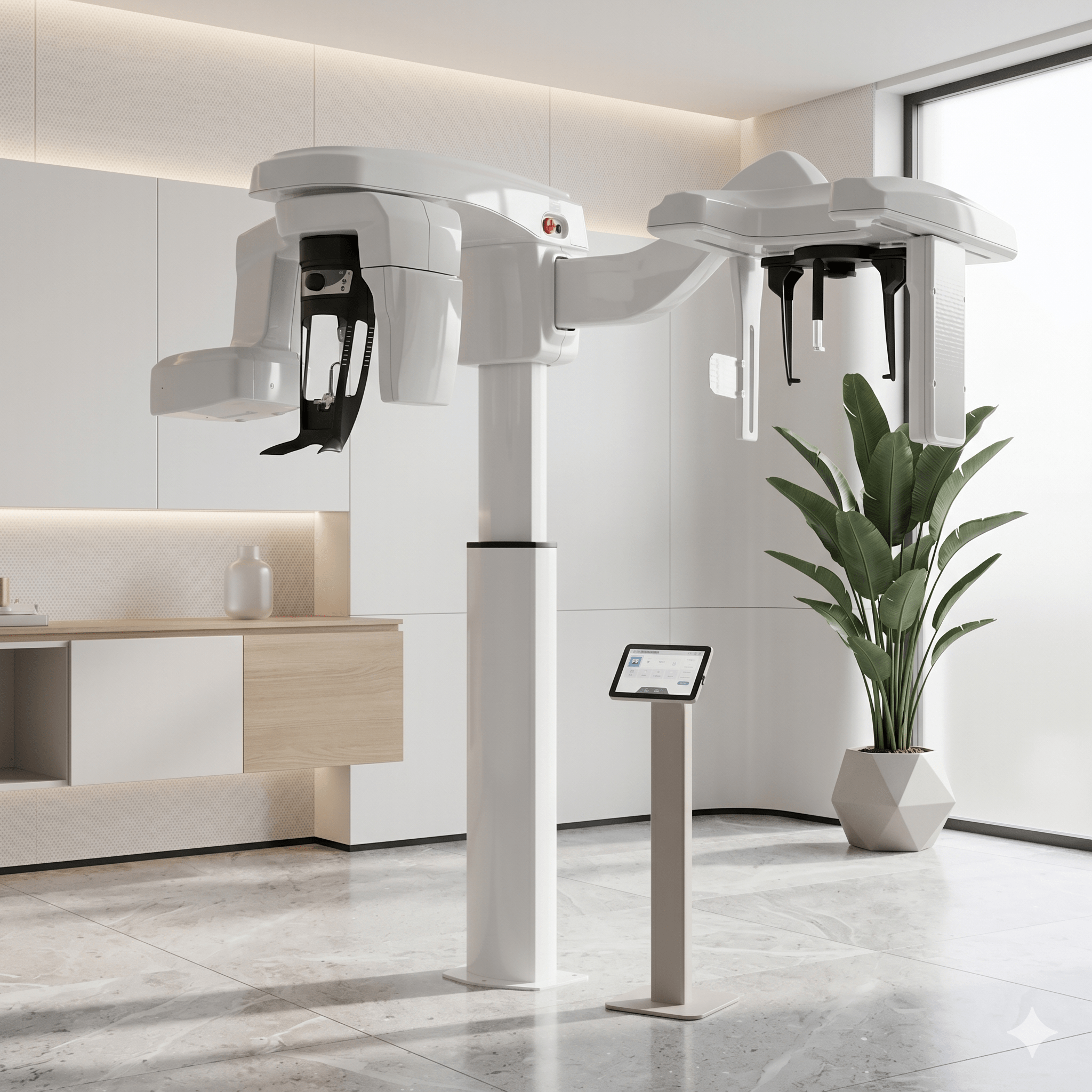
More precise diagnosis
Imaging studies help provide a clearer view of dental and maxillofacial structures for a more complete evaluation.
2D and 3D studies
We offer radiographic and tomographic alternatives according to the treatment needs and the required diagnosis.
Supports treatment planning
Images help guide different dental procedures with more clinical information.
Diagnostic center
What studies do we perform at our imaging center?
We offer radiographic and tomographic studies that help obtain a more complete view of dental and maxillofacial structures to support different dental treatments.
2D
2D radiographic studies
We perform studies such as panoramic, cephalometric, periapical, and bitewing radiographs, depending on the diagnostic need and the type of evaluation required.
3D
Cone Beam CT scan
Cone Beam 3D CT provides a more detailed view of anatomical structures, supporting the planning of cases that require greater diagnostic precision.
BENEFITS
What can a dental imaging center provide?
A properly indicated imaging study can help improve clinical evaluation, guide diagnosis, and support better treatment planning.
P
Greater diagnostic precision
Images allow us to observe in greater detail areas that cannot always be fully evaluated through clinical examination alone.
T
Better planning
Studies help guide treatments in a more informed way, with a clearer view of the structures involved.
R
Faster response
Having imaging studies in the same environment can facilitate the diagnostic process and move toward a more complete evaluation.
C
Support for different specialties
2D and 3D studies can be useful in orthodontics, implants, oral surgery, endodontics, and other dental treatments.
Better treatment starts with a better diagnosis
If you need information about radiographs, CT scans, or studies for a more complete dental evaluation, message us on WhatsApp and we will guide you according to the type of study you need.
Request information Call nowTypes of studies
Imaging studies that may be part of a dental evaluation
Depending on the case and the treating specialty, different 2D or 3D studies may be requested to support diagnosis and clinical planning.
RX
Panoramic radiograph
Provides a general view of both dental arches, dental structures, and jaw areas in a single image.
CF
Cephalometric radiograph
It is often used as support in orthodontic studies and in the evaluation of growth and craniofacial relationship.
BW
Bitewing radiograph
Helps observe interproximal details and may be useful in detecting cavities between teeth and evaluating restorations.
CP
Periapical radiograph
It may be indicated as support in growth evaluations, especially in certain orthodontic cases.
CB
3D Cone Beam CT
Provides a more detailed three-dimensional visualization for cases that require a deeper anatomical analysis.
Study selection
Not all cases require the same type of image
The choice of study depends on the reason for consultation, the treatment to be planned, and the level of diagnostic detail needed by the professional.
01
Evaluation according to clinical need
The same patient may require different studies depending on whether the case is related to orthodontics, surgery, implants, endodontics, or another specialty.
02
Support for a more precise treatment plan
Choosing the appropriate study helps obtain more useful information to guide clinical decisions with better diagnostic support.
The right study can make a difference in diagnosis
If you need guidance on dental radiographs or CT scans, message us and we will help you identify which type of study may correspond to your case.
Ask on WhatsApp Call nowWhen may it be needed?
Imaging studies that support different dental treatments
Depending on the case, the professional may request radiographic or tomographic studies to obtain a more complete evaluation before defining the treatment.
1
Orthodontics
Studies such as panoramic, cephalometric, or periapical radiographs may be part of orthodontic growth evaluation and planning.
2
Implants and surgery
A 3D CT scan may be useful when a more detailed view of bone and anatomical structures is required.
3
Endodontics
Some studies help evaluate compromised teeth and complement clinical analysis in root canal treatments.
4
General evaluations
Images may be part of the initial diagnosis when a more complete review of oral health is needed.
Diagnostic confidence
An appropriate study can provide more clarity for diagnosis
Having supporting images helps observe certain structures in greater detail and better guide clinical decisions in different dental treatments.
★★★★★
They guided me on which study I needed, and that helped me move forward faster with the evaluation of my treatment.
Dental diagnosis patient
★★★★★
Having the study done in the same dental setting made everything more organized. They explained what it was for and why it was important in my case.
Dental planning patient
★★★★★
The CT scan helped a lot in the evaluation of my case. I felt the diagnosis was much clearer and more complete.
3D CT scan patient
A better image can lead to better planning
If you need information about dental radiographs or CT scans, message us and we will guide you on the type of study that may correspond to your needs.
Ask on WhatsApp Call nowFREQUENTLY ASKED QUESTIONS
We answer common questions about our imaging center
These are some common questions from patients who need radiographs or CT scans as part of their dental evaluation.
What is the difference between a 2D study and a 3D CT scan?
2D studies provide flat radiographic images, while a 3D CT scan provides a more detailed volumetric view, useful in cases that require greater diagnostic precision.
Do all patients need a CT scan?
No. The need for the study depends on the case and on what the treating professional needs to evaluate in order to guide diagnosis and planning.
What studies may be requested for orthodontics?
Depending on the case, panoramic and cephalometric radiographs may be requested, and in some cases a periapical study may also be part of the orthodontic evaluation.
Is a panoramic radiograph useful for a general evaluation?
Yes. It is a commonly used study to obtain a broad view of both arches, teeth, and related structures in a single image.
Can I ask for guidance on which study I need?
Yes. We can help guide you according to your dentist’s indication or the type of dental evaluation you want to have.
Take the first step toward a more precise evaluation
If you need information about panoramic, cephalometric, bitewing, periapical, or 3D CT studies, message us on WhatsApp and we will help guide you.
Ask on WhatsApp Call now